
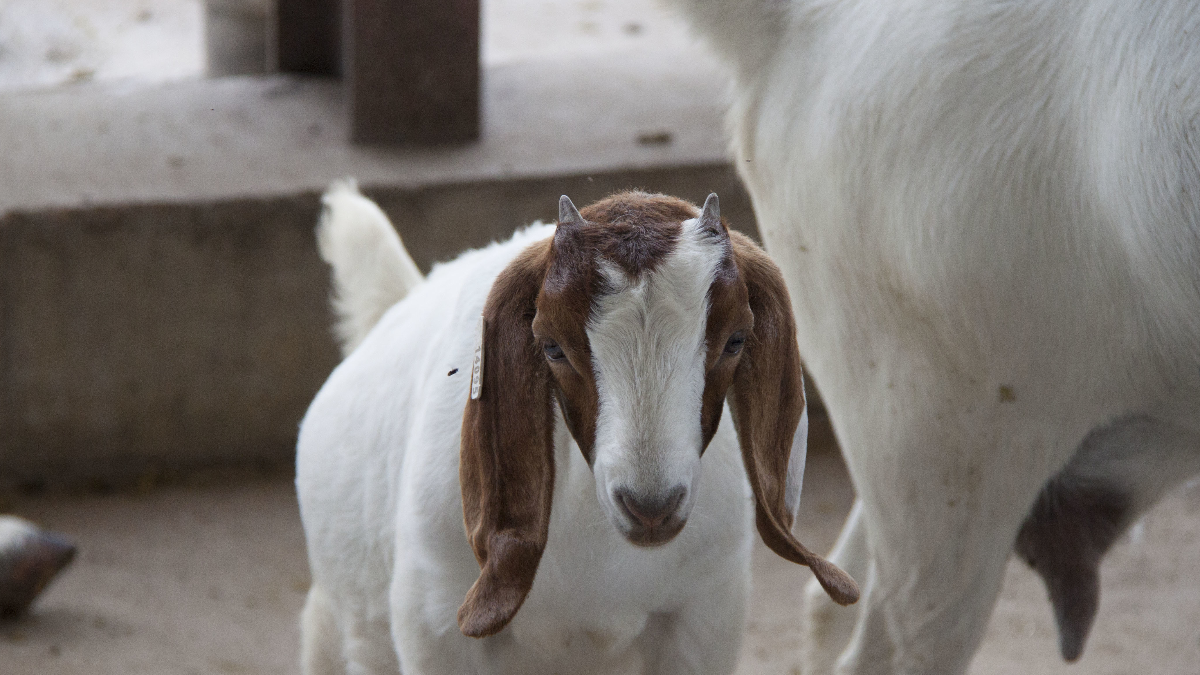
May 20 – The International Goat Research Center at Prairie View A&M University continues to lead the way in animal research through its innovative discovery in all aspects of better developing agriculture for the future. A team of Cooperative Agricultural Research Center (CARC) researchers under the direction of the Animal Systems Research Scientist Leader, Dr. Gary Newton, came together to study how the expression of the fucosyltransferase gene in goats effects pregnancy, ultimately contributing the breeding of these animals. A comprehensive article about their findings was recently published in Theriogenology: Agricultural Journal of Animal Reproduction.

Abstract:
Regulation of the expression of the alpha(1,2)fucosyltransferase (FUT) genes and their enzymatic products, including the H-type 1 antigen (HT1), on the luminal surface of the uterus is believed to be critical for establishment of pregnancy in mammals. The FUT1 gene is a marker for conception rates in dairy cows and HT1 is a marker for uterine receptivity in rodents. To determine the spatiotemporal expression patterns of FUT1 and FUT2 genes in goats, endometrial tissues were obtained on six days spanning the estrous cycle (Days 5, 11, 13, 15, 17 and 19) and seven days spanning early pregnancy (Days 5, 11, 13, 15, 17, 19 and 25). In all data, we found no effect of status (cyclic or pregnant; P > 0.1) and pooled data where appropriate. We cloned FUT1 cDNA from goat endometrium and made probes from it for Northern and slot blot analyses. The analyses indicated that FUT1 gene expression was high until Day 13, and then declined. In situ hybridization revealed a change in the cell-specificity of FUT1 gene expression over the estrous cycle and early pregnancy. In situ hybridization signal intensity scores indicated that FUT1 expression by uterine epithelium was high on Day 5, moderate on Day 11, and minimal on subsequent days. In situ hybridization signals in uterine glandular epithelial cells remained high from Day 5 to Day 13, with weaker signals thereafter. Quantitative reverse transcription-PCR (RT-qPCR) assays were used for quantitation of FUT1 and FUT2 mRNAs. Quantitative RT-qPCR data were generated from endometrium collected from cyclic and pregnant animals on Days 5, 11 and 17. Relative levels of FUT1 mRNA were high on Days 5 and 11, but then fell 5-fold by Day 17 (P < 0.01). FUT2 mRNA concentrations were below the accurate detectable limit of the assay. High levels of HT1 were observed on the apical surface of uterine luminal epithelia on Days 5, 15, 17 and 19, with much lower levels on Days 11 and 13. Thus, data suggests that FUT1 is the primary enzyme responsible for the high levels of HT1 antigen present on the uterine luminal epithelium between Days 5 and 11 of the estrous cycle and early pregnancy. But changes in the expression of the FUT1 gene does not directly correlate to HT1 staining, which increased from Day 13–15. Future studies are required to understand the regulation of the HT1 antigen on the luminal surface of endometrium.
To read the entire article, please visit the journal website here.
This work was supported by the USDA National Institute of Food and Agriculture, 1890 Extension Formula Program projects under Section 1444.

Taelor Smith
Communications Specialist
tasmith@pvamu.edu
(936) 261-5155